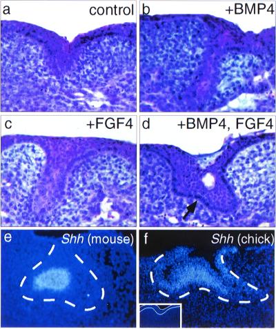
Figure 3.
Induction of chick oral epithelial appendages by BMP and/or FGF. (a) Section through a control, untreated chick mandible after 6 days of culture showing region of thickened epithelium. (b and c) Bud-like structures induced in chick mandibles after 6 days of culture with 100 ng/ml of exogenous BMP4 (b) or FGF4 (c). (d) More advanced epithelial structure induced to form in chick mandibles after 6 days of culture with BMP4 and FGF4 (100 ng/ml each). Note convoluted epithelium (arrow). The clear space is a cyst. (e) Localization of Shh transcripts in the enamel knot of an E14.5 mouse molar tooth germ. (f) Shh expression induced in the central portion of the epithelial structure by addition of BMP4 and FGF4 to chick mandibles in explant culture. The dotted line in e and f indicates the location of the basal lamina separating epithelium and mesenchyme. (Inset) Shh is not expressed in control explants; the epithelium resides between the white lines.

