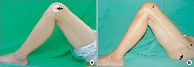
Fig. 1.
(A) Lateral epicondyle of the femur was marked with an electrocardiogram lead (arrow). (B) Two kinds of palpable anatomical landmarks - anterior margin (a) of the greater trochanter (arrow) and lateral epicondyle of the femur (a') - were identified and the line aa' was defined as the "palpable mechanical axis" of the femur in the sagittal plane.

