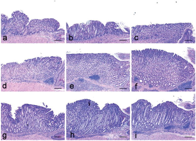
Figure 2.
Gastric histopathology of H. pylori infection in gulo−/− with low vitamin C supplementation (a, d, g) or high vitamin C supplementation (b, e, h) versus C57BL/6 (WT) controls (c, f, i). (a–c) No significant lesions developed in uninfected mice in any group at 32 weeks. (d–f) Equivalent gastric lesions developed in all H. pylori-infected groups at 16 WPI, although gulo−/− mice with low vitamin C supplementation (d) exhibited a trend for less severe epithelial defects, foveolar hyperplasia and dysplasia. (g–i) Equivalent gastric inflammation developed in all groups at 32 WPI, although gulo−/− mice with low vitamin C supplementation (g) had less severe epithelial defects, mucous metaplasia and foveolar hyperplasia.

