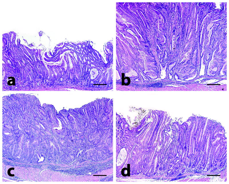
Figure 1.
Gastric histology in mice. At 32-34 weeks old, uninfected male mice exhibited spontaneous gastric epithelial dysplasia with inflammation (a). H. pylori infection significantly increased inflammation, oxyntic atrophy, foveolar hyperplasia, and dysplasia in the corpus of age-matched male mice at 28 (WPI) (b). Sulindac treatment exacerbated inflammation but not hyperplasia or dysplasia in H. pylori-infected mice (c). H. pylori antimicrobial therapy and sulindac significantly reduced inflammation, oxyntic atrophy, foveolar hyperplasia, and dysplasia (d). Tissues were stained with H&E; bar = 400 μM.

