Figure 6.
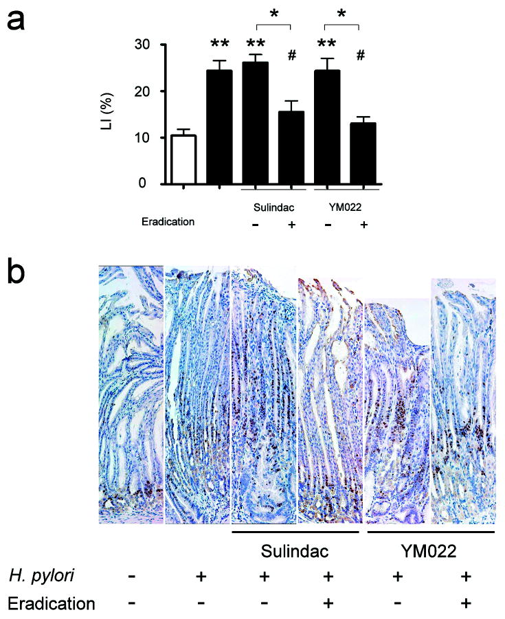
Cell proliferation in gastric mucosa; Ki-67 labeling indices (LI) (b), and immunohistochemical staining of Ki-67 (c). Ki-67 LI were compared in corpus mucosa (b). H. pylori infection resulted in significantly higher Ki-67 LI in mice (p<0.01). Sulindac or YM022 did not alter H. pylori-associated increase of Ki-67 LI in INS-GAS mice. H. pylori-infected mice receiving the combination of sulindac or YM022 and H. pylori eradication had significantly lower Ki-67 LI compared to untreated, infected mice (both p<0.05) or infected mice that received sulindac or YM022 (p<0.05, respectively). Proliferating cells were positively stained for Ki-67 (c). In helicobacter-uninfected INS-GAS mice, proliferating cells were restricted to the isthmus regions. Zone of proliferating cells expanded from isthmus regions to foveolar regions in the untreated, H. pylori-infected mice and infected mice that received sulindac or YM022. Proliferating cells were restricted to the isthmus regions irrespective of the hyperplastic foveolar glands in the infected mice that received a combination of sulindac or YM022 and H. pylori eradication. White bar, H. pylori-uninfected mice; black bars, H. pylori-infected mice; Ki-67 positive cells were stained in brown color. (Compared to uninfected, untreated mice or comparison between indicated groups: *, p<0.05; **, p<0.01; ***, p<0.001. Compared to infected mice: #, p<0.05).

