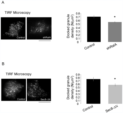
Figure 4. Effect of RalA silencing on insulin granule docking.
A) Experiments were performed under TIRF illumination. INS-1E cells were co-transfected with a plasmid encoding IAPP-EGFP (a fluorescently labeled protein targeted to insulin granules) and either an empty vector (control) or shRalA. On the left: representative TIRF images of control and shRalA transfected cells. On the right: histograms depict the number of granules/µm2 counted in the “footprint” of each cell using TIRF illumination in control conditions and in cells transfected with shRalA. Results are the means ± SEM (n = 23 cells control; n = 40 cells shRalA). * p<0.05 one way ANOVA. Scale bar: 12 µm B) INS-1E cells were co-transfected with a plasmid encoding NPY-mRFP (a fluorescently labeled protein targeted to insulin granules) and either an empty vector (control) or a plasmid driving the expression of a dominant-negative form of Sec8 (Sec8-ΔΝ). Quantitative PCR analysis confirmed that in transfected cells the dominant-negative transcript was approximately 1000-folds more abundant than the endogenous Sec8 mRNA. On the left: representative TIRF images of control and Sec8-ΔΝ transfected cells. On the right: histograms represent number of granules/µm2 counted in the “footprint” of each cell under TIRF illumination in control conditions and in cells transfected with ΔSec8. Results are mean ± SEM (n = 22 cells control; n = 29 cells, Sec8-ΔΝ). * p<0.05 one-way ANOVA. Scale bar: 12 µm.

