
Figure 1.
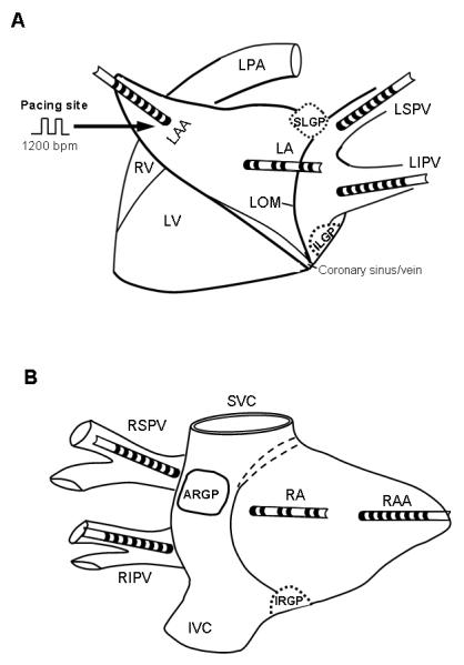
Schematic representation and catheter position in the atrium. (A) Left thoracotomy approach. SLGP: superior left ganglionated plexi (GP), located adjacent to the junction of left superior pulmonary vein (LSPV) and left atrium (LA); ILGP: inferior left GP located near the junction of left inferior pulmonary vein (LIPV) and LA. Multi-electrode catheters were sutured to the LSPV, LIPV, LA and left atrial appendage (LAA). Continuous rapid pacing (1200 bpm) was performed at the LAA. LV: left ventricle; RV: right ventricle; LOM: ligament of Marshall. (B) Right thoracotomy approach. ARGP: anterior right GP, located adjacent to the right superior pulmonary vein (RSPV) - atrial junction; IRGP: inferior right GP, located at the junction of the inferior vena cava (IVC) and both atria. SVC: superior vena cava. Similarly, multi-electrode catheters were sutured to RSPV, right inferior pulmonary vein (RIPV), right atrium (RA) and right atrial appendage (RAA).