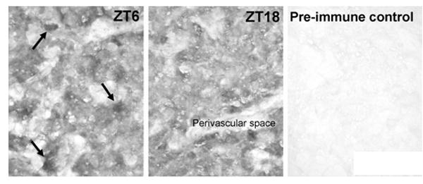
Fig. 3. Immunohistochemical detection of Mbnl2 protein in the rat pineal gland.

Animals were housed in a controlled lighting environment for two weeks (LD12:12). Pineal glands were obtained at ZT6 and ZT18. Immunohistochemical detection was done with the polyclonal rabbit anti-Mbnl2 used in Fig. 2B. at a dilution of 1:1,000. A pre-immunization negative control (ZT18) is displayed. Note the universal staining of the cells in the pineal parenchyma and a few intensively stained cells (arrows in ZT6). Scale bar, 50 μm. For further details see the Materials and Methods section.
