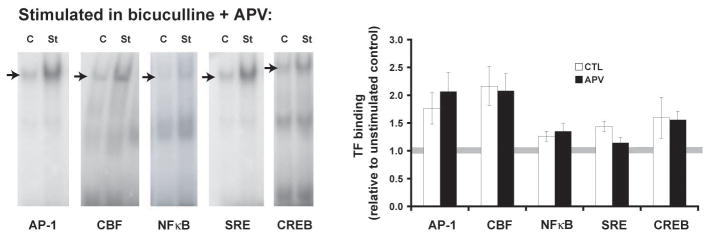
Fig. 1.
Stimulation-induced transcription factor binding does not require NMDA receptors (or LTP) if action potentials are maintained. To insure that action potentials were maintained during NMDA receptor antagonist exposure (50 μM D-APV), synaptic stimulation was delivered in a theta burst pattern of stimulation (TBS) with bicuculline [7]. Hippocampal CA1 mini-slices were sampled 5 minutes after stimulation. Nuclear protein extracts were assessed for transcription factor binding by electrophoretic mobility shift assays (EMSAs). Example gels are shown on the left, with arrows marking the bands representing specific binding. Plotted on the right are data from 8–11 (control) and 7–8 (APV) separate EMSAs. Transcription factor binding in APV-treated slices are not significantly different from the slices without the drug. Binding to SRE is not affected by APV at this time point (p=0.04, a=0.01).

