
Figure 20.
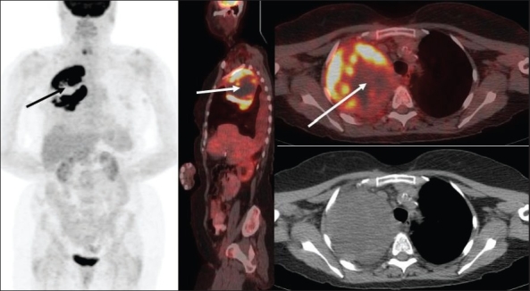
Axial fused PET/CT (top right) and CT (bottom right) images at the level of the upper lungs, with a sagittal fused PET/CT image (middle) and an MIP image (on left), from a PET study of a 71-year-old lady with a large right upper lobe mass. Bronchoscopic evaluation and two CT-guided biopsies failed to reveal malignancy. PET scan clearly shows a large malignant right upper lung mass with a large ‘cold’ central necrotic area (arrows) and intense FDG uptake peripherally. A third CT-guided biopsy from the periphery of the mass based on the PET findings confirmed NSCLC