Figure 2.
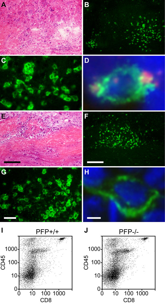
CD8+ cells infiltrate the spinal cord in chronically demyelinated mice irrespective of perforin expression. Spinal cord sections were prepared from perforin-competent (A–D) and perforin-deficient (E–H) mice at 180 dpi. (A–H) Representative inflammatory lesions in white matter are shown by hematoxylin and eosin stain (A, E). The lesions contain many cells that are immunopositive for CD8 in both perforin-competent (B, C) and perforin-deficient mice (F, G). Perforin co-staining (red) was observed in CD8+ cells (green) within demyelinated lesions in perforin-competent mice (D) but not in perforin-deficient mice (H); blue indicates DAPI. (I, J) Flow cytometric analysis at 180 dpi confirmed that the number of CD45hiCD8+ cells present within isolated spinal cord infiltrating leukocytes did not differ between perforin-competent (I) and perforin-deficient mice (J). Scale bar in E = 50 µm and also refers to A. Scale bar in F = 50 µm and refers to B. Scale bar in G = 10 µm and refers to C. Scale bar in H = 1 µm and refers to D. dpi = days post-infection

