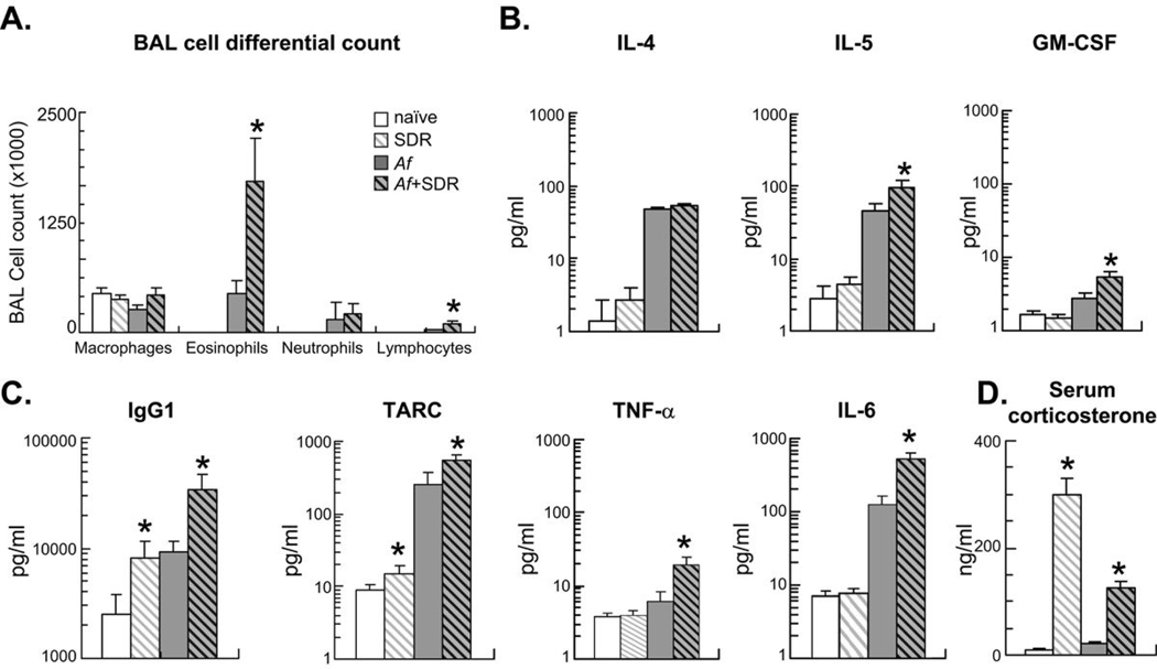
Figure 2.
Mice were sensitized (i.p.) and challenged (i.n.) with Af extract and were also exposed to social stress (SDR) daily between days 7–12 as indicated. Sensitized mice were studied 48 h after a single Af challenge (on day 15). Naïve and SDR mice were also studied on day 15. (A): BAL differential cell count was evaluated in Giemsa preparations. Stress and allergic sensitization enhanced numbers of eosinophils and lymphocytes but not neutrophils and macrophages in the BAL fluid. There was no difference between naïve and SDR mice. (B–C): Chemokine, cytokine and immunoglobulin levels in the BAL were measured in the same groups by SearchLight technology. Increased eosinophilia was paralleled by enhanced levels of the eosinopoietic IL-5 and GM-CSF (B), increased levels of the B-cell derived IgG1 and the innate immune cell-derived TARC, TNF-α and IL-6 (C). (D): Stress significantly increased serum corticosterone levels p<0.01: SDR vs. naïve, Af or Af+SDR. Blood for endocrine measures was taken on day 15. (A–D): (Mean±SEM of n=8 per group) *p<0.05 Naïve vs. SDR and Af vs. Af+SDR; Repeated measure ANOVA followed by Tukey's HSD.

