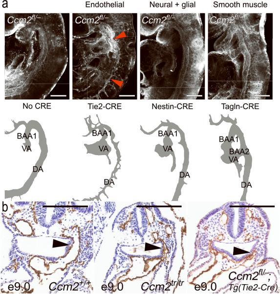
Figure 2. Vascular defects are endothelial autonomous.
(a) Whole mount immunofluorescence demonstrates normal, uniform caliber branchial arch arteries and aortae in all Ccm2fl/- embryos except the endothelial (Tie2-CRE) mutant, which has an irregular, narrow lumen (red arrowheads). Cartoons below are provided for orientation. BAA1 = first branchial arch artery, BAA2 = second branchial arch artery, VA = ventral aorta (or aortic sac), DA = dorsal aorta. (b) The narrow branchial arch arteries are well demonstrated on paraffin sections taken at E9.0. As opposed to the wild type embryo (left panel) the first branchial arch artery (arrowhead) is similarly narrowed and irregular in both the complete knockout (Ccm2tr/tr, middle panel) and the endothelial mutant (Ccm2fl/-;Tg(Tie2-CRE), right panel). Scale bars: 200 μm.

