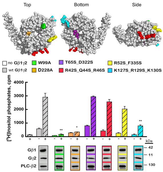
Fig. 5. Regions of Gβ involved in PLC-β2 activation.
Groups of residues mutated on the Gβ surface [1got.pdb] are colored and coordinate with colors in the bar graph and associated blots. Wild-type and mutant Gβ1 subunits were tested in the absence (-) or presence (+) of PLC-β2 for their ability to activate PLC-β2 (*p<0.01 **p<0.005, error bars represent standard error). Immunoblot analysis confirmed equal expression of all proteins utilized.

