Figure 1.
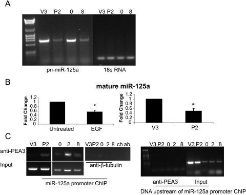
EGFR signaling through PEA3 represses miR-125a expression. (A) RT-PCR for pri-miR-125a (lanes 2–5) and 18s RNA (lanes 6–9) on vector (V3), PEA3 (P2), OVCA433 untreated (0), and OVCA433 + 8 hours of EGF treatment (8). (B) TaqMan PCR for mature miR-125a on untreated OVCA433 cells or treated with EGF for 8 hours and in V3 and P2 cells. Expression was normalized to untreated OVCA433 and V3 cells, respectively. *P < .05. (C) ChIP for PEA3 association with the PEA3 binding sites immediately upstream of miR-125a in V3 and P2 cells. ChIP was also performed on OVCA433 cells untreated or treated with EGF for 2 or 8 hours. IP with anti-β-tubulin was used as a negative control. ChIP performed in the absence of chromatin (ch) or antibody (ab) did not result in PCR products. Input chromatin for each sample is shown. ChIP was also performed on chromosomal DNA upstream of the miR-125a promoter on samples and input DNA.

