
Fig. 1.
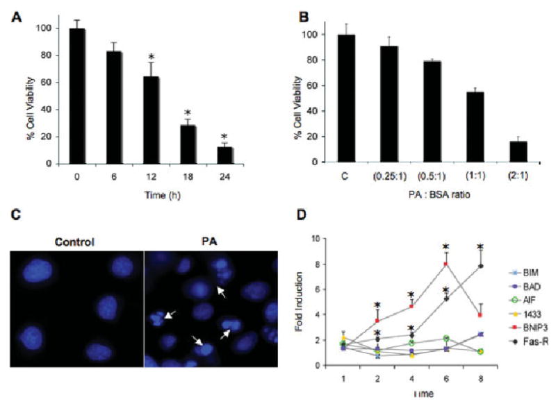
Palmitic acid-induced lipotoxicity in NGFDPC12 cells. A: NGFDPC12 cells were exposed to PA/BSA (2:1 ratio), and viability was determined using WST-1 assay at the indicated times. B: NGFDPC12 cells were exposed to different PA/BSA ratios (0.25:1, 0.5:1, 1:1, and 2:1 with BSA at a concentration of 0.150 mM) for 24 hr. Cell viability was measured by the WST-1 assay. C: NGFDPC12 cells were exposed to 0.150 mM BSA without PA (control) or PA/BSA (2:1; PA) for 12 hr. Cells were stained with Hoechst (10 ng/liter), and nuclei were visualized under fluorescent microscopy. Arrows indicate nuclei showing chromatin condensation and fragmentation. D: Regulation of apoptosis-asociated genes in NGFDPC12 cultures exposed to PA/BSA (2:1). Quantitative RT-PCR experiments show the time-dependent mRNA expression of Bim, Bad, AIF, BNIP-3, 14.3.3, and FAS-R. GAPDH mRNA expression was used as the internal control. Data represent mean 6 SEM of three independent experiments. ★P < 0.05 compared with control.