
Fig. 5.
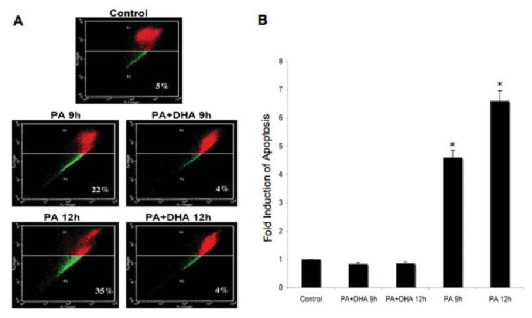
DHA reduces mitochondrial depolarization of NGFDPC12 cells undergoing PA-induced lipotoxicity. A: NGFDPC12 cells were treated with PA/BSA alone (PA) or in the presence DHA (PA + DHA) for 9 or 12 hr, and mitochondrial depolarization was determined by JC-1 flow cytometry assays. Representative flow cytometric plots are shown. B: Quantification of mitochondria depolarization (R2 in JC-1 flow cytometry) in NGFDPC12 cells undergoing PA-induced lipotoxicity. Data represent mean ± SEM of three independent experiments. ★P < 0.05.