
Figure 2.
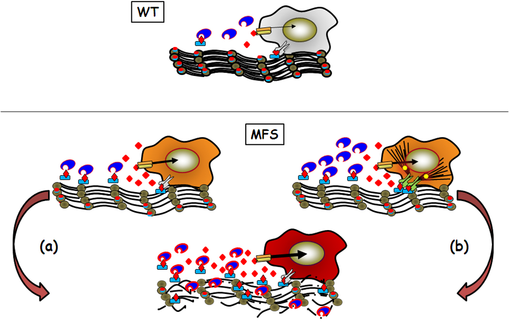
Model of normal regulation of TGFβ by microfibrils (WT) and perturbations associated with microfibril deficiency in Marfan syndrome (MFS). Two models of MFS pathogenesis leading to loss of tissue integrity (bottom) are shown. In the first model (a), physiological levels of activators (proteases (blue indented ovals) and integrins (grey symbols)) drive disease by increasing release of TGFβ (red diamonds) from matrix-free LLCs (blue rectangle with red diamond). In the second model (b), cells enhance TGFβ activity by producing more proteases, by increasing integrin-mediated LLC activation (also through heightened cytoskeletal tension (black lines)), and by stimulating stress-response signaling pathways (yellow stars). Although the two models focus on the onset of MFS pathogenesis, impaired LLC sequestration (model (a)) could be the immediate trigger of cellular responses to an abnormal matrix (model (b)).