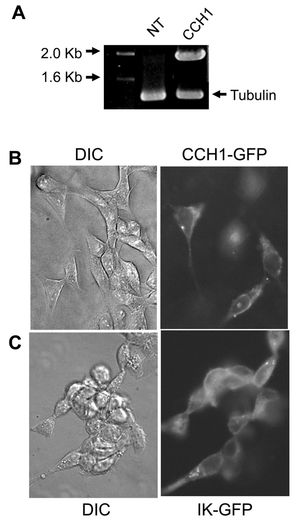
Figure 4.
The functional expression of CCH1 in HEK293 cells. A) Reverse transcriptase (RT) PCR analysis revealed the presence of CCH1 transcripts in HEK293 cells that had been transfected with CCH1-GFP plasmid DNA. CCH1 transcripts were not detected in non-transfected HEK293 cells. Tubulin is shown as a loading control. Total RNA was isolated from HEK293 cells by standard protocols and used as template in the RT-PCR reaction. PCR amplified products were separated by agarose electrophoresis and visualized by ethidium bromide. HEK293 cells were transiently transfected with CCH1-GFP plasmid DNA as follows: approximately 1µg plasmid DNA was added to 100 µl, of Opti-MEM I reduced serum media and mixed with 1.75 µl of Lipotectamine 2000 and the mixture was incubated for 30 min at room temperature. HEK293 cells were maintained in Dulbecco’s Modified Eagle medium (DMEM) supplemented with 4 mM L-Glutamine, 10% fetal bovine serum at 37ºC and 5% CO2. β) Localization studies revealed a predominant plasma membrane distribution of Cch1-GFP in HEK293 cells, similar to the localization pattern of a well-characterized plasma membrane-bound IK+ channel shown here as a plasma membrane marker, (DIC, differential interference contrast) (C). HEK293 cells were fixed in 4% paraformaldehyde for 20 min at room temperature and washed in PBS. The fluorescence label (GFP) was visualized by using filters S484/15 for excitation and S517/30 for emission.

