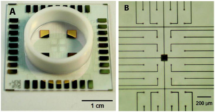
Figure 1.

Photographs illustrating features of microelectrode array. (A) A lower magnification imagei llustrating the entire MEA including peripheral contacts, Teflon ring for developing culture space and larger reference electrodes. (B) A higher magnification image illustrating electrode distribution and conductor leads.
