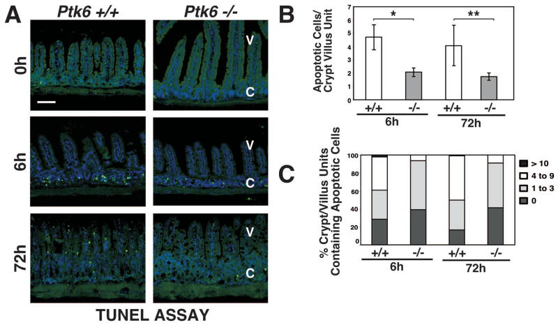
Figure 2. DNA-damage induced apoptosis is compromised in the absence of PTK6.
(A) Radiation–induced apoptosis was examined in the small intestine from untreated and γ-irradiated Ptk6 +/+ and Ptk6 −/− mice at 0, 6 and 72 hours post irradiation using the TUNEL assay. Labeled apoptotic cells were detected with Avidin-FITC (apoptotic cells stain green). The size bar represents 100 μm. Quantification was performed and is presented as a histogram (B) and frequency histogram (C) of apoptotic cells per crypt-villus unit. (B) At least 50 crypt-villus units per section were scored. Values shown are mean ± S.D. from two sections from at least three different mice per group. *P-value = 0.026; **P-value = 0.032. V: villi; C: crypts.

