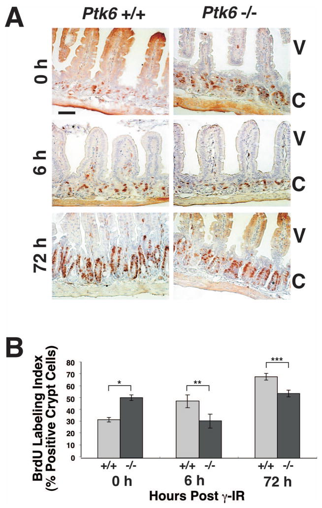
Figure 7. Increased numbers of intestinal crypt epithelial cells proliferate in wild-type mice than Ptk6 null mice in response to DNA damage.
(A) Proliferation was analyzed by examining BrdU incorporation in sections of small intestine from untreated and γ-irradiated Ptk6 +/+ and Ptk6 −/− mice at 0, 6 and 72 hours post irradiation. S-phase cells were pulse-labeled with BrdU for 1 hour before animals were sacrificed. BrdU incorporation was detected with antibodies against BrdU and DAB (brown). Positive nuclei are detected in the crypts, while some diffuse background staining is seen in the upper villus. V denotes villi and C crypts. Untreated Ptk6 −/− mice exhibit an extended zone of proliferation at 0 h, but proliferation levels are higher in wild-type mice at 6 and 72 h post irradiation. (B) The percent of BrdU positive cells per crypt in wild-type (+/+) and Ptk6 null (−/−) mice at 0, 6, and 72 h post irradiation is shown. Bars, +/− SD. (*P-value = 0.015; **P-value = 0.023; ***P-value = 0.047).

