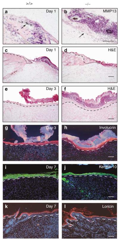
Figure 2. MMP13 expression analysis and microscopic assessment of wounds in MMP13-deficient mice.
(a, b) In situ hybridization for MMP13 on wounds of (a) mmp13 >/> controls and (b) mmp13 −/− mice followed by H&E staining to detect transcripts on sections of the left-hand site of wounds 1 day post injury. The arrow points to keratinocytes of the leading wound edge. Bar: 50μm. (c–f) H&E-stained sections of the left-hand site of wounds (c, d) 1 day and (e, f) 3 days after injury. The dashed line indicates basement membrane. The migrating tip is marked by the dotted line. Bar: 100μm. (g, h) Full re-epithelialization of mmp13 −/− and control wounds, as demonstrated by wound sections taken 3 days post injury and immunostained for involucrin (red signal). Nuclei were counterstained with Hoechst dye (blue signal), which also stained the scab. The basement membrane is indicated by a dashed line. Bar: 100μm. (i–l) Sections of wounds taken 7 days post excision were immunostained for the differentiation marker (i, j, green signal) keratin-10 and (k, l, red signal) loricrin to demonstrate complete keratinocyte differentiation. Nuclei were counterstained with Hoechst dye (blue signal). The dashed line indicates basement membrane. Bars: 100μm.

