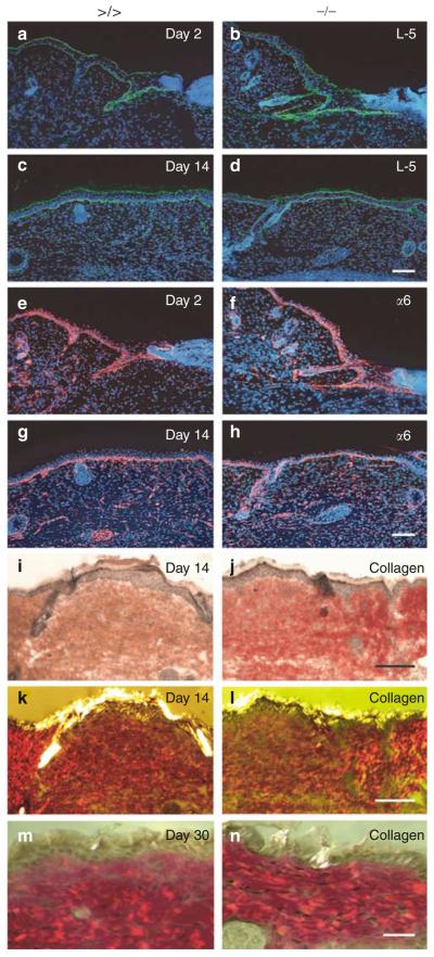
Figure 4. Immunofluorescence analyses of basement membrane re-organization and visualization of dermal collagen fibers in wounds of mmp13 −/− mice.
(a–h) Laminin-5 (a–d, green signal) and (e–h, red signal) α6-integrin were visualized by immunofluorescence on sections of mmp13 >/> (left panel) and mmp13 −/− (right panel) wounds analyzed 2 and 14 days after injury. Nuclei were counterstained with Hoechst dye (blue signal). Bars: 100μm. (i–n) To visualize collagen fibers of wounds taken at (i–l, bar: 50μm) day 14 and (m, n, bar: 30μm) day 30, sections were stained by Picrosirius red and documented by both (i, j) bright field and (k–n) crossed polarization.

