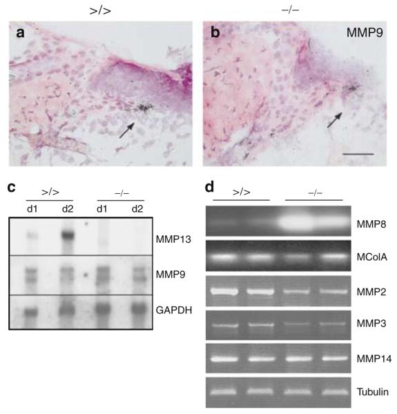
Figure 5. MMP expression analyses in the early phase of wound healing.
(a, b) In situ hybridization followed by H&E staining to detect MMP9 transcripts on sections of the left-hand site of wounds from (a) mmp13 >/> and (b) mmp13 −/− mice. The arrow points to keratinocytes of the leading wound edge. Bar: 50μm. (c) Northern blot analyses were performed with total RNA prepared from day 1 and 2 wounds of mmp13 >/> and mmp13 −/− mice and hybridized to MMP13 and MMP9 cDNA. Quality and quantity of the RNA was confirmed by visualization of GAPDH. (d) Semiquantitative PCR of different MMPs performed on cDNA prepared on total RNA isolated from day 1 wounds of mmp13 >/> and mmp13 −/− mice. After gel electrophoresis, PCR products of MMP8 and McolA were blotted and hybridized to the corresponding PCR fragments. The autoradiograph was scanned and reverted. β-Tubulin was used as an internal standard.

