Figure 5.
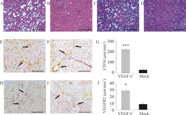
Effect of VEGF-C overexpression on angiogenesis of orthotopic tumors. H&E-staining of representative PC-3/VEGF-C (A, orthotopic; C, subcutaneous) tumors and PC-3/mock (B, orthotopic; D, subcutaneous) tumors. PC-3/VEGF-C tumors showed angiogenic morphology with a rich network of capillaries compared with PC-3/mock tumors. There were significantly more blood capillaries (CD34 positive, arrows) in the PC-3/VEGF-C tumors (E, 220 ± 15 μm/mm2, n = 29) compared with PC-3/mock tumors (F and G, 37 ± 6 μm/mm2, n = 24), p < 0.001. Density of VEGFR2-positive capillaries was analyzed similarly. Again there were significantly more VEGFR2-positive capillaries in the PC-3/VEGF-C tumors (H, 30 ± 0.3 μm/mm2, n = 29) than in the PC-3/mock tumors (I and J, 9 ± 0.1 μm/mm2, n = 24), p < 0.05. Bar 500 μm.

