
Fig. 2.
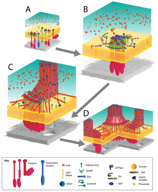
Schematic view of signaling pathways that lead to actin organization at invadopodia or podosomes. (A) At the initial stage of adhesion formation, integrins or other unidentified receptors bind to components of the ECM (grey), leading to clustering of receptors into PtdIns(4,5)P2-enriched areas of plasma membrane. (B) Recruitment of Src to adhesion sites leads to phosphorylation of several proteins such as cortactin, WASP, FAK and regulators of small GTPases. Continuous actin nucleation relies on the continuous and strong activation of the Arp2/3 complex at the membrane through the synergistic action of cortactin and WASP-family proteins. (C) DRF/mDia1 elongates actin filaments into columnar structures from the branched actin network that was previously induced by N-WASP, the Arp2/3 complex and cortactin. (D) Podosomes or invadopodia are mechanically connected through a network of radial actin filaments that lie parallel to the substratum.