
Fig. 3.
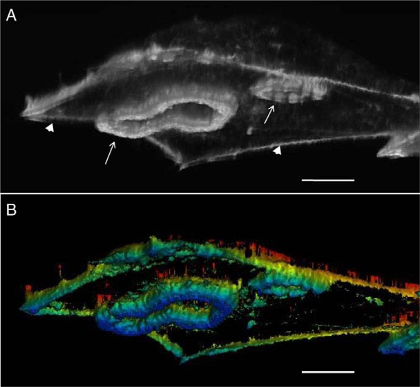
Three-dimensional (3D) reconstruction of F-actin structure in a BHKRSV cell. (A) A 3D reconstruction was derived by combining images from confocal planes viewed from the side of the basal (adherent) face. F-actin staining was carried out after fixation in 4% paraformaldehyde with TRITCphalloidin. 3D reconstruction and rendering of the actin cytoskeleton was carried out through EDIT3D software, using grey-level images of each confocal z-stack (developed by Yves Usson and Franck Parazza, UMR CNRS 5525, Grenoble, France). Actin stress fibers are indicated by arrowheads, and the collective organization of podosomes and invadopodia by arrows. (B) A color scale was added, purely to indicate the relative position of the z-plane. The most basal plane was colored blue. Scale bars: 5 μm.