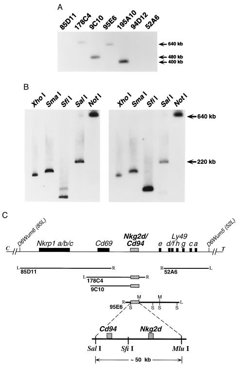
Figure 4.
Mapping of murine Nkg2d and Cd94 within the NKC. (A) Southern blot analysis of YACs previously shown to form a contig spanning an ≈2.0-megabase region of the murine NKC on chromosome 6 (39), schematically represented in C. This blot was hybridized with both mCD94 (shown) and mNKG2-D (not shown) full-length cDNA probes; identical patterns were observed. (B) Southern blot analysis of YAC 95E6 digested with the indicated enzymes and hybridized with mNKG2-D (Left) or mCD94 (Right) cDNA probes. The murine Nkg2d and Cd94 loci are discriminated on SfiI digestion of YAC 95E6. (C) Schematic representation of the murine Nkg2d and Cd94 loci (hatched boxes) positioned between Cd69 and the Ly49 cluster (solid boxes). The positions of each YAC previously aligned within the NKC contig are shown below (solid lines). Where known, the right (R) and left (L) ends of each YAC are indicated. SalI (S) and Mlu I (M) sites present in YAC 95E6 are shown for reference. An expanded region of YAC 95E6 representing the ≈50-kb SalI–Mlu I fragment containing both loci is shown at the bottom. The exact position of the SfiI site separating the Cd94 and Nkg2d loci and the precise location of the individual genes on the fragment have not been determined.

