
Fig. 5.
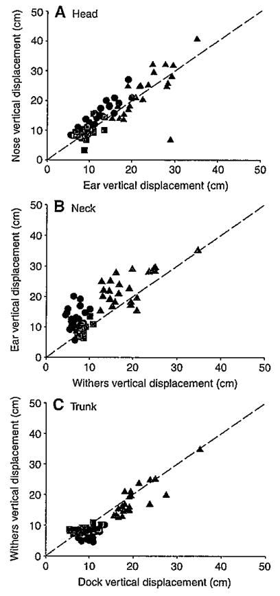
Vertical translations of the head (A), neck (B) and trunk (C) during walks (black circles), trots (red squares) and canters (blue triangles). Translation of each segment is depicted by plotting the vertical displacement of its rostral landmark (y-axis) against that of its caudal landmark (x-axis). Broken lines have a slope of 1 indicating vertical segmental translation with no rotation.