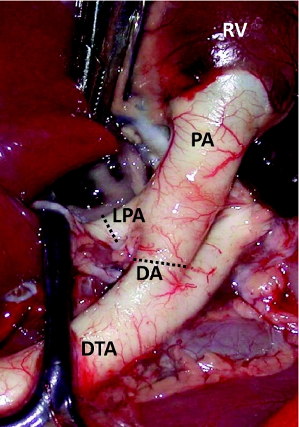
Figure 1. An image from a fetal sheep demonstrating the anatomy of the ductus arteriosus (DA) in relation to the main pulmonary artery (PA), the left pulmonary artery (LPA), the right ventricle (RV) and descending thoracic aorta (DTA).
Flow probes were placed around the LPA and the DA, at the positions indicated by the dotted lines, for the simultaneous measurement of blood flow in the LPA and DA, respectively.

