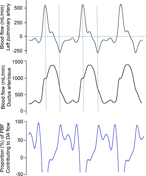
Figure 3.
Instantaneous measures of blood flow in the left pulmonary artery (top panel) and ductus arteriosus (middle panel) acquired over 3 consecutive cardiac cycles from a fetal sheep. The bottom panel shows the percentage of total pulmonary blood flow (PBF) contributing to right-to-left (from pulmonary into systemic circulation) flow of blood through the ductus arteriosus. Negative PBF values (top panel) indicate retrograde flow of blood away from the lungs.

