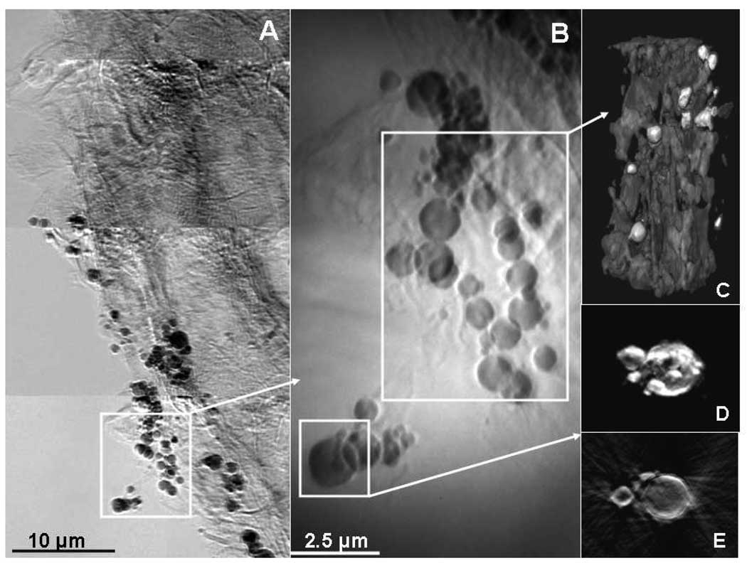
Figure 1.
TXM mosaic image of S. foliosa roots taken at 9 keV in absorption contrast shows dark particles and dark channels due to absorption by Hg (A). Blowup (B) shows greater detail. 2D stills from tomography of particles from (B) show particles with greatest absorption (lightest), possibly surrounded by biofilms (C). 2D tomographic still (D) and slice (E) of large particle indicate that highest Hg concentrations (lightest intensity) are on the outside of the fairly hollow particles.

