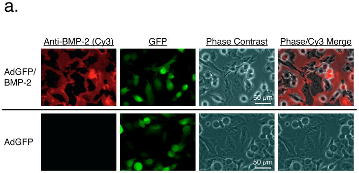
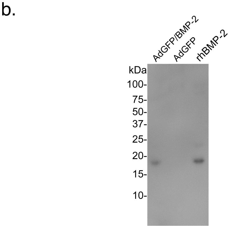
Figure 1. Adenoviral co-expression of BMP-2 and GFP.
a. Immunofluorescence detection of BMP-2 and GFP in adenovirus transduced R3230 cells. R3230 cells infected with AdGFP/BMP-2 virus (top) and control AdGFP viruses (bottom) 48 hours previously were assessed for expression of BMP-2 (left column) and GFP (2nd column). A phase contrast (3rd column) and merge of BMP-2 and phase contrast images (right column) are also shown.
b. Secretion of BMP-2 by adenovirus transduced R3230 cells. The equivalent of 75 μL of cell culture supernatant collected between 24 and 48 hours from R3230 cells transduced with AdGFP/BMP-2 (left lane) or control GFP viruses (middle lane), and 50 ng of recombinant human BMP-2 (rhBMP-2; right lane), were resolved on a 16% Tris-Tricine SDS-PAGE gel and subjected to immunoblot analysis using a BMP-2-specific antibody.


