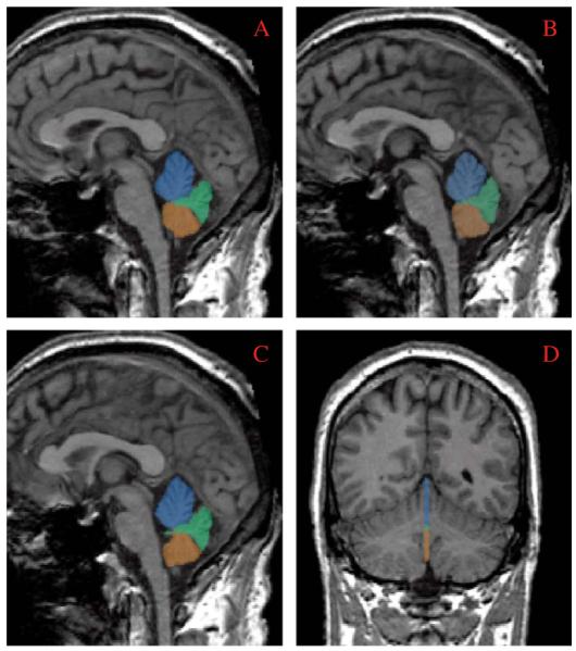
Fig. 1.

Panes A-C demonstrate manual tracings performed on the three sagittal slices of the cerebellar vermis, including a mid-sagittal (B) and 2 parasagittal slices (A and C). Pane D demonstrates all three slices in a coronal view. Parcellated areas included anterior superior vermis (lobules I-V) appearing in blue; posterior superior vermis (lobules VI-VII) in green: and inferior posterior vermis (lobules VIII-X) in orange. See text for a description of the anatomic criteria used for the parcellations.
