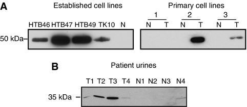
Figure 2.
Western blot validation of cathepsin D. (A) Samples of CM (5 μg load in each case) were probed for cathepsin D. Expression was seen in all established cell lines examined but absent in one normal sample included for comparison purposes. Increased expression by tumour cultures in two out of three primary tumour (T)/normal (N) matched pairs was also apparent. Parallel Coomassie staining was performed for loading control purposes. (B) Urine from four healthy controls and four patients with RCC were probed for cathepsin D. 15 μl of urine was loaded in each case. Uncropped blots are presented in Supplementary Figure 2.

