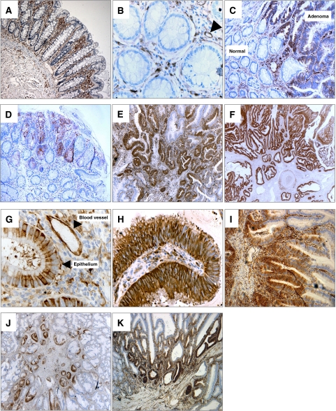
Figure 1.
Fascin is overexpressed in colorectal adenomas: immunoreactivity correlates with tumour size, histology, and dysplasia and is focussed towards the stalk. Fascin immunoreactivity was detected in specimens of human colorectal adenomas by immunoperoxidase staining. (A and B) Normal colorectal mucosa showing positive staining in the stroma (arrowheads indicates endothelial cells in B), × 100 and × 400 magnifications, respectively. (C) The contrast in fascin immunoreactivity between a large tubulovillous adenoma with severe dysplasia and the adjacent non-neoplastic tissue (labelled), × 100 magnification. (D, E and F) Representative figures showing fascin immunoreactivity in a small tubular (D, × 100), medium tubulovillous (E, × 50) and large tubulovillous (F, × 25) adenoma. (G and H) Higher magnification images to show the subcellular distribution of fascin immunoreactivity within the adenoma epithelium: G, a medium tubulovillous adenoma with mild dyspplasia, × 400; H, medium tubular adenoma with moderate dysplasia, × 400 (arrowheads, endothelial cells in G). (I, J and K) Images showing the focal localisation of fascin expression around the adenoma stalk (I, medium tubulovillous adenoma, × 100; J, medium tubular adenoma, × 25; K, large tubulovillous adenoma, × 50).

