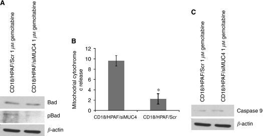
Figure 3.
Assessment of intrinsic apoptotic pathway in CD18/HPAF/Scr and CD18/HPAF/siMUC4 cells in response to gemcitabine treatment. (A) Cells were seeded in 10 cm petridishes and treated with 1 μM gemcitabine for 48 h as described in methodology. A total of 50 μg protein from each cell extract was resolved by SDS–PAGE (15%), followed by immunobloting with anti-pBad, anti-Bad and anti-β-actin (internal control) antibodies. pBad protein level was more in CD18/HPAF/Scr cells compared with CD18/HPAF/siMUC4 cells. (B) Cytosolic fractions were prepared from 1 μM gemcitabine-treated CD18/HPAF/Scr and CD18/HPAF/siMUC4 cells. The amount of cytochrome c protein from each fraction was then measured with a commercially available cytochrome c ELISA kit. Level of cytochrome c in the cytosol of CD18/HPAF/siMUC4 was more compared with CD18/HPAF/Scr cells. (C) A total of 20 μg protein from each cell lines treated with 1 μM gemcitabine for 48 h was resolved by SDS–PAGE (10%), followed by immunobloting with antibodies against cleaved caspase-9 and β-actin (internal control). CD18/HPAF/siMUC4 cells showed more cleaved caspase-9 compared with CD18/HPAF/Scr cells. These findings indicate up-regulation of intrinsic apoptotic pathway in CD18/HPAF/Scr cells.

