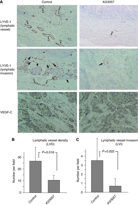
Figure 3.
Immunohistochemical expression of LYVE-1 and VEGF-C in orthotopic tumours. (A) Orthotopically planted OCUM-2MLN cells frequently invade into lymphatic vessels. The number of lymphatic vessels at the invasive edge of tumours was fewer in mice treated by Ki23057, compared with the control. The number of lymphatic invasion was fewer in the gastric tumours treated by Ki23057, compared with the control. Lymphatic vessels located at the invasive edge of tumours were often enlarged and dilated. Expression level of VEGF-C was not different between the two groups. The LYVE-1 antibody stains lymphatic vessels (arrowheads) and highlights lymphatic invasion (arrows). Many lymphatic vessels were found at the invasive edge of tumours. The LYVE-1 antibody-stained vessels devoid of red blood cells corresponded to lymph vessels, whereas blood vessels were not stained (asterisks). Single-stained endothelial cells were excluded. (B) Lymphatic vessel density (LVD) was determined by LYVE-1-positive vessels without cancer emboli. Respective mean LVD in control and Ki23057 mice respectively were 26.5 and 10.4. The mean LVD was significantly decreased by Ki23057. (C) Lymphatic vessel endothelial hyaluronan receptor-1 immunostaining highlighted the presence of lymphatic invasion. Lymphatic vessel invasion was identified as the presence of cancer emboli within the channels lined by LYVE-1-positive vessels. Respective mean LVI (lymphatic vessel invasion) in control and Ki23057 mice were 3.5 and 0.7. The LVI was significantly decreased by Ki23057. Lymphatic vessel invasion or LVD was determined by counting the number of LYVE-1-positive vessels with or without cancer emboli in four high-power fields ( × 100) in areas of the highest lymphatic vessel as ‘hot spots’.

