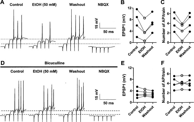
Fig 5. EtOH inhibits PN EPSPs and AP firing evoked by stimulation of granule cell axons.
A. Sample traces of current-clamp recordings from a PN in which a train of five stimuli (100 Hz) delivered in the molecular layer induced non-NMDA EPSPs that triggered a burst of action potentials. Bath application of EtOH (50 mM) reversibly decreased the peak amplitude of the EPSPs and reduced action potential number (these data were digitized at 5 kHz, which explains the variability in AP amplitude). The non-NMDA EPSPs were fully blocked by NBQX (20 μM). B and C. Summary graphs illustrating the inhibitory effect of EtOH (50 mM) on EPSP1 amplitude and action potential number. Analysis by repeated measures ANOVA followed by Tukey's posthoc test revealed that EtOH significantly decreased EPSP1 amplitude (p < 0.01 vs. control and p < 0.05 vs. washout) and the number of action potentials/train (p < 0.001 vs. both control and washout) (n = 4). D. Same as in A but in the continuous presence of bicuculline (20 μM); the stimulation intensity was decreased to obtain a comparable number of action potential/train as in the absence of bicuculline. Note that EtOH neither decreased EPSP1 amplitude nor action potential number in presence of this agent. E and F. Summary graphs illustrating the lack of an effect of EtOH (50 mM) on EPSP1 and action potential number in presence of bicuculline (n = 5).

