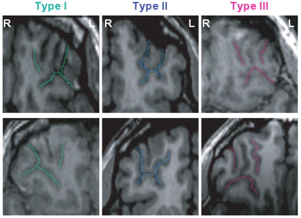
Fig. 2.

MRI images of major three types of ‘H-shaped’ sulcus. Examples of the major three sulcogyral patterns from six different subjects. On the axial plane of SPGR (spoiled gradient-recalled images), sulci of Type I, II, III are delineated with green, blue and pink colour, respectively. Upper and lower columns demonstrate left and right hemisphere. At this level, olfactory sulcus cannot be observed in most cases. Sulcal continuities of the medial and lateral orbital sulci were determined by evaluating several consecutive axial slices rather than just a single slice. L, left; R, right.
