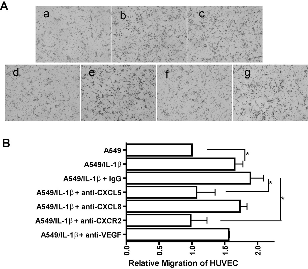
Figure 3. IL-1β significantly augments the angiogenic activity of NSCLC by inducing the expression of angiogenic CXC chemokine genes.
A, CM was collected from A549 cells treated with either control medium (a) or medium containing 2.5 ng/ml of IL-1β (b) for 24 h and CM from IL-1β-treated A549 cells was preincubated with 10 µg/mL of non-immune IgG (c) or an anti-CXCL5 monoclonal antibody (d) or an anti-CXCL8 monoclonal antibody (e) or an anti-CXCR2 monoclonal antibody (f) or an anti-VEGF monoclonal antibody (g) at 37°C for 1 h. A migration assay was performed by stimulating HUVECs with the indicated CM as described in Materials and Methods. Migrated dells were photographed after 16 h of incubation at 37°C. B, Image analysis of HUVECs migration was quantified using the ImageJ software program. Data were expressed as the mean ± SE. *, p<0.05.

