
Figure 2.
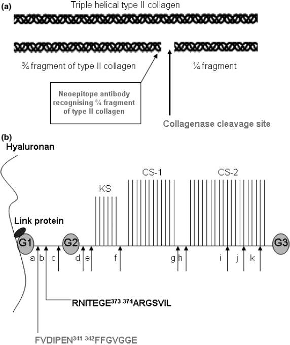
(a) Structure of triple helical collagen and cleavage by collagenases. Schematic representation of fibrillar type II collagen is shown. The 3/4; and ¼. fragments resulting from the cleavage of a triple helical collagen molecule by collagenases and the detection site of neoepitope antibodies is also shown. (b) Structure of aggrecan and cleavage by aggrecanases and MMPs. Schematic drawing showing the structure of aggrecan. G1 – 1st globular domain, G2 – 2nd globular domain, G3 – 3rd globular domain, KS – keratan sulphate glycosaminoglycan (GAG) attachment region, CS-1 – 1st chondroitin sulphate GAG region, CS-2 – 2nd chondroitin sulphate attachment region. Hyaluronan can bind numerous aggrecan molecules to form a large polymeric structure. The interaction of aggrecan with hyaluronan is strengthened by link protein. The reported cleavage sites of aggrecan by MMPs (a, c, d, e, f, g) and aggrecanases (b, h, i, j, k) are shown. The cleavage sites most relevant to OA are in the interglobular domain and the specific amino acid residues involved in cleavage are shown (adapted from Nagase & Kashiwagi 2003).