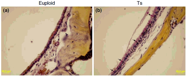
Figure 4.

Representative figures to visualize goblet cells in the middle ear mucosae of Ts65Dn (Ts) mice and wild-type controls (euploid). Few goblet cells could be found in the middle ear mucosa in the control mouse (a). By contrast, the goblet cells were present at high density among other cells in the epithelium of the middle ear cavity of Ts65Dn mouse [(b) typical goblet cells are indicated by arrows]. Goblet cells have a distinctly polarized morphology in which the nucleus stains black in colour at the cell bas, and the mucus stains a deep rose colour in the middle and apical portions of the cell. Scale bars = 20 μm.
