
Figure 1.
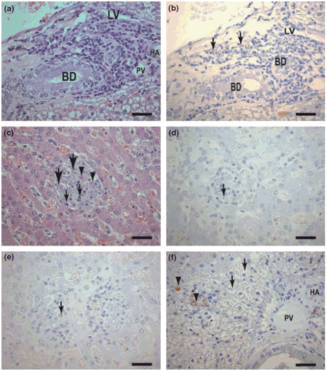
(a–f) Liver sections of dogs naturally infected with L. (L) chagasi. (a,b) Asymptomatic dog: (a) Portal space with a cellular infiltrated of plasma cells lymphocytes and macrophages. HE (Bar = 16 μm). (b) Same field showing immunolabelled amastigotes forms of Leishmania in macrophages (large arrows). Streptoavidin-peroxidase method (Bar = 16 μm). BD, bile duct; LV, lymphatic vessel; HA, hepatic arteriole; (c) Asymptomatic dog. Observe an intralobular granulomas formation comprising macrophages (epithelioid cells) (large arrows), plasma cells (arrowheads) and lymphocytes (small arrows). HE (Bars = 16 μm). (d) Same field showing immunolabelled amastigotes into granulomas macrophages (arrows), Streptoavidin-peroxidase method (Bar = 16 μm). (e) Symptomatic dog: Observe two confluent granulomas in the centre of the figure. Amastigote could be seen (arrow). (f) Symptomatic dog: Kupffer cells intensely parasitized (arrowheads). Note an intense swelling of the hepatocytes (‘balloon cells’) (arrows) (Bar = 16 μm). BD, bile duct; LV, lymphatic vessel; HA, hepatic arteriole; PV, portal vein.