
Figure 2.
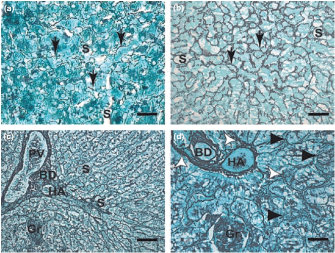
(a–d): Liver sections of control dogs and naturally infected dogs with L. (L) chagasi. (a,b,c.d): (a) Control dog: Higher magnification showing a delicate network of intralobular collagen fibres (reticular fibres) (arrows). Gomori ammoniacal silver-staining (Bars = 16 μm). Note collagen fibres extend through sinusoids. (b) Symptomatic dog: Intralobular fibrosis characterized by collagen fibres extend through sinusoids. Note conspicuous collagen thickening in the space of Disse HE (Bars = 16 μm); (c,d) Symptomatic dog: (c) Lower magnification (panoramic view) showing an intense proliferation of collagen and reticulin fibres detected by ammoniacal silver-staining extend through portal spce (Bars = 32 μm). (d) Detail showing hepatic cells that had become isolated from the sinusoidal blood by the fibropoiesis (arrowheads). Observe dense and coiled fibres (white arrows) extend through sinusoids from the portal tract. Gomori ammoniacal silver-staining (Bars = 16 μm). BD, bile duct; HA, hepatic arteriole; PV, portal vein; S, sinusoids; Gr, granuloma.