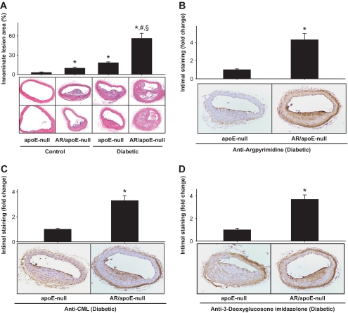
FIG. 7.
Genetic ablation of aldose reductase exacerbates diabetic lesion formation and AGE accumulation. A: Photomicrographs of cross sections of innominate arteries of 20-week-old nondiabetic (control) and diabetic apoE-null and akr1b3-apoE–null mice. Sections were stained with hematoxylin and eosin, and the lesion area was quantified by image analysis. Data are presented as means ± SE. *P < 0.01 vs. apoE-null (control), #P < 0.01 vs. aldose reductase/apoE–null (control) and §P < 0.01 vs. apoE-null (diabetic). Arterial sections of diabetic apoE-null and akr1b3-apoE–null mice stained with anti-argpyrimidine (B), anti-CML (C), and anti–3-deoxyglucosone imidazolone (D) antibodies. The extent of staining was quantified by image analysis. Data are presented as means ± SE. *P < 0.05 vs. apoE-null (diabetic). AR, aldose reductase. (A high-quality color digital representation of this figure is available in the online issue.)

