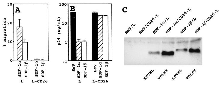
Figure 4.
Inactivation of SDF-1α by specific cleavage of CD26/DPPIV. (A) Tenfold-diluted culture supernatants of clone C3 (L) or B5 (L-CD26) infected with SeV/SDF-1α or SeV/SDF-1β were assayed for the level of chemotactic activity. (B) MT4 cells were treated with tenfold-diluted culture supernatants of clone C3 or B5 infected with the wild-type SeV (SeV), SeV/SDF-1α (SDF-1α), or SeV/SDF-1β (SDF-1β) and then infected with NL43. The levels of p24 antigen production 4 days after infection are shown. (C) Proteins in 250 μl of culture supernatant of clone C3 or B5 infected with the wild-type SeV, SeV/SDF-1α, or SeV/SDF-1β were precipitated with ethanol and analyzed by Western blotting using anti-SDF-1 antiserum. The N-terminal amino acid sequence of each protein band is shown.

