
Fig. 1.
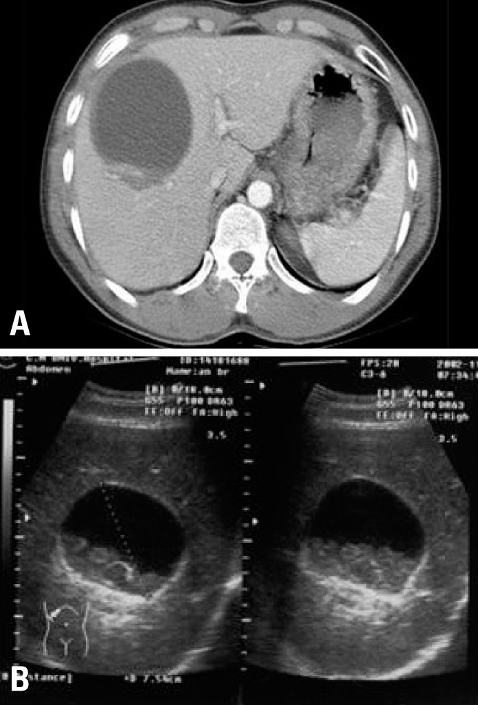
(A) Computed tomography of the abdomen with contrast medium showed a low-density cystic mass, measuring 9×8 cm, in the right hepatic lobe. (B) Ultrasonography of the liver showed a lesion with the characteristic appearance of a hydatid cyst with a hyperechogenic capsule and echogenic material.