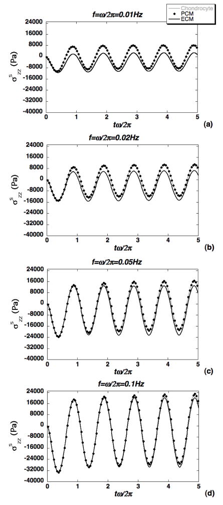
Figure 4.
Illustration of force transmission at three points along the symmetry axis located at 90% of the cell radius (chondrocyte), 90% of the PCM thickness and at the top of the microscale domain (ECM). Axial solid stress is shown at four loading frequencies: (a) f=0.01Hz, (b) f=0.02Hz, (c) f=0.05Hz and (d) f=0.1Hz.

